Osseointegration / Osseofixation: What Are the Differences?
1. Large Differences Are Found in the Field of Medical Contraindications and in the Field of Burdens and Risks for the Patients
| Method of Osseointegration | Method of Osseofixation | |
|---|---|---|
| Permanent medical contra-indications for oral implant treatment which will lead to de-selection of the patient by the treatment provider | Unfavorable medical conditions (diabetics, hypertension, various medications, IV bisphosphonate treatment, etc.) Smoking Insufficient bone supply and unfavorable conditions for bone augmentation |
n.A. |
| Temporary medical contra-indications for oral implant treatment which will lead to temporary postponing of the patient by the treatment provider | IV bisphosphonate treatment Periodontal infections, cysts in the bone, infections in the bone, recent radiation therapy |
IV bisphosphonate treatment, recent radiation therapy |
| Reasons for patient’s refusal to undergo oral implant treatment | Long duration of treatment Very high costs of implant treatment High risks associated with bone augmentation Additional costs of bone augmentation Fear of repeated pain during multi-step surgical protocols Unwillingness to wear an intermediate removeable denture or to be without teeth for some time Fear of experiencing periimplantitis which will lead to pain, infections and eventually to the loss of large amounts of bone and loss of the implants |
Despite the comparatively lower treatment costs, some patients will still postpone treatment for financial reasons |
Table 1: The table shows major differences between the Method of Osseointegration and the Method of Osseofixation regarding permanent and temporary contra-indication as well as regarding patient’s reason(s) for not accepting the treatment.
2. Different Regions for Anchorage
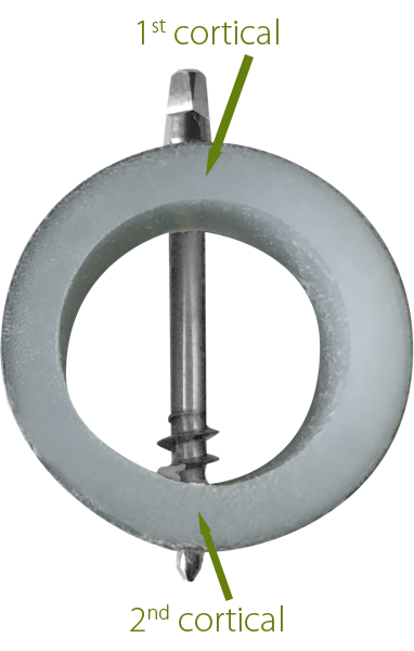
After the removal of teeth, the jaw bones collapse to some extend - a process which is called "atrophy". We know today that oral implants will not prevent this collapse. Conventional oral implants (Method of Osseointegration) will however promote the collapse and lead to more bone loss. The reasons are: due to their large diameter, their rough surfaces and their multi-piece design, they trigger in many cases the loss of the bone in the area of the 1st cortical. This creates then a chronic infection and finally leads to the loss of the osseointegrated implant. Often patients request (after lengthy periods of suffering) simply the removal of the affected implants.
The Older Method of "Osseointegration"
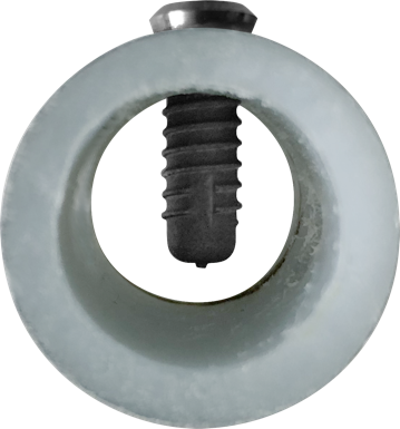
A conventional "osseointegrated" implant is anchored in the unstable 1st cortical and therefore any vertical bone loss may trigger unfavourable developements like a peri-implantitis.

The New Method of Osseofixation
Implants which are designed for osseofixation are anchored in the 2nd cortical. Peri-Implantitis does not occur because the shafts of the implants are thin and polished. The load transmission to the bone takes place in the area of the 2nd cortical. The principle of "bicortical" anchorage was taken over from the field of traumatology, where it is used since approximately 1975. This type of anchorage allows to work in immediate functional leading protocols.

3. No More Periimplantitis
Therapy with 2-stage implants (using the Method of Osseointegration) is quite complicated. It takes a long time and often several surgical interventions. But this is not the main problem: many of these implants which have been "successfully" placed and are equipped with crowns and bridges, develop after two to three years an insidious and malicious disease named "periimplantitis".
The disease starts with a small inflammation around these implants, followed by ever larger infections. The disease progresses in fits and starts. Over time, most of the jaw bone around the implants is dissolved by the infection. This disease is found only around the old type of implants and we know today that the reason is their large diameter, their rough surface and their construction principle in general. The danger that this disease poses is underestimated and often downplayed.
For us it is not understandable why dentists still use such implants - but many do this!
Around the Strategic Implant® (if it was placed correctly by the treatment provider and maintained well by the patient), peri-implantitis will not develop. This was shown in all studies which were published so far[1]. This alone is an unbeatable advantage over all other methods of oral implant placement and it increases the safety of the patients drastically (compared to the old Method of Osseointegration).
[1] Dobrinin O., Lazarov A, Konstantinovic V.K., et al. Immediate-functional loading concept with one-piece implants (BECES/BECES N /KOS/ BOI) in the mandible and maxilla- a multi-center retrospective clinical study. J. Evolution Med. Dent. Sci. 2019;8(05):306-315, DOI: https://doi.org/10.14260/jemds/2019/67
Pałka Ł, Lazarov A. Immediately loaded bicortical implants inserted in fresh extraction and healed sites in patients with and without a history of periodontal disease. Ann Maxillofac Surg 2019;9:371-8.
Lazarov A. Immediate functional loading: Results for the concept of the Strategic Implant®. Ann Maxillofac Surg 2019;9:78-88.
Gosai H., Anchilla Sonal, Kiran Patel, Utsav Bhatt, Phillip Chaudhari, Nisha Grag. Versatility of Basal Cortical Screw Implants with Immediate Functional Loading J. Maxillofac. Oral. Surg. 2021, https://doi.org/10.1007/s12663-021-01638-6
Comparison Between the Strategic Implant® Treatment and Treatments Using the Older Method of Osseointegration
| Strategic Implant® | Stage | Conventional |
|---|---|---|
| Inspection Diagnostic procedures Treatment plan |
1 | Inspection Diagnostic procedures Treatment plan |
| Preparation of the oral cavity: removal of teeth, roots and cysts. Implant placement, impression taking for the lab, the control of X-ray diagnostics | 2 | Preparation of the oral cavity: removal of teeth, roots and cysts, waiting time for healing after the beforementioned treatments |
| Trying of the bridge frame (0 - 1 days after implant placement) | 3 | Bone augmentation and sinus-lifting (if there are indications) |
| Delivery of bridge (1 - 3 days after implant placement) | 4 | 1 - 2 months after implant placement, adequate bone healing provided |
| — | 5 | Healing of the bone after tooth extraction (2 - 4 months) |
| — | 6 | Placement of gingiva former |
| — | 7 | Impression taking |
| — | 8 | Fixing of the bridge |
| Treatment duration: 1 - 5 days Number of appointments: 4 - 5 |
Total | Treatment duration: 4 - 24 months Number of appointments: 8 - 12 |
Control appointments are necessary after 3 months and then after every 12 – 24 months (according to the decision of the treatment provider).
